MRI (Open)
Our Open MRI is 3 sided open. Patient don't feel any kind of fear or discomfort during the test. People who get nervous in small places (claustrophobic) may feel better in open MRI. An open MRI is easier to use for people who are very overweight. But not all medical centers have this kind of MRI machine. The open MRI scanner is designed to provide optimal image quality comparable to traditional closed or tunnel MRI scanners. The scanner offers flexibility of positioning to put patients at ease whilst having their MRI scan. 96% of patients who are unable to be scanned in a conventional scanner have been successfully scanned in the Open MRI Scanner.

CT Scan
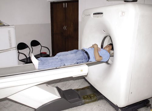
CT scan, is an X-ray procedure that combines many X-ray images with the aid of a computer to generate cross-sectional views and, if needed, three-dimensional images of the internal organs and structures of the body. A CT scan is used to define normal and abnormal structures in the body and/or assist in procedures by helping to accurately guide the placement of instruments or treatments. Our CT Scan offers patients low-dose CT scans. CT scan is used by the radiologists at Sobti Hospital to guide procedures. The images generated during the CT scan are viewed on a computer monitor and printed on film. They can also be transferred to a CD or DVD if the patient demands.

Digital X-Rays
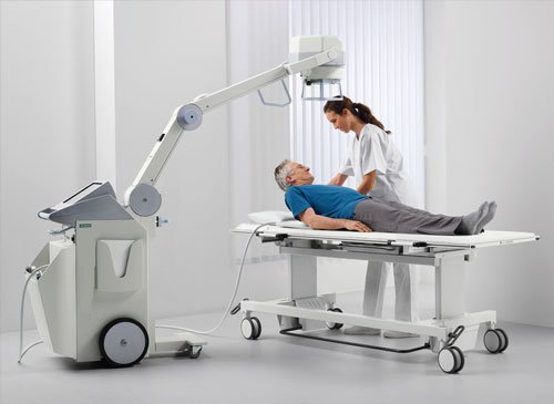
Sobti Hospital does Digital radiography which is a form of X-ray imaging, where digital X-ray sensors are used instead of traditional photographic film. Digital X-Rays is replacement of the former analog methods of detection, with the almost instantaneous development of images on a digital display, instead of the former methods of film and the associated delay in time and chemistry consumption. Advantages include time efficiency through bypassing chemical processing and the ability to digitally transfer and enhance images. Also less radiation can be used to produce an image of similar contrast to conventional radiography.

Doppler
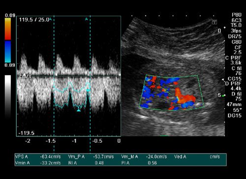
Doppler ultrasound is a noninvasive test that can be used to estimate your blood flow through blood vessels. This test may be done as an alternative to more invasive procedures, such as arteriography and venography, which involve injecting dye into the blood vessels so that they show up clearly on X-ray images. It is very helpful to diagnose blood clots, poorly functioning valves in your leg veins, a blocked artery, decreased blood circulation into your legs (peripheral artery disease), narrowing of an artery, such as in your neck (carotid artery stenosis) esp. in patients coming with stroke in the hospital.
